Minimally Invasive Spine Surgery
- Home
- Minimally Invasive Spine Surgery
Minimally Invasive Spine Surgery
What is minimally invasive surgery?
Though minimally invasive spine surgery was first performed in the 1980s, it has recently seen rapid advances. Traditional, open back surgeries can potentially damage normal uninvolved musculature. There are potential chances for damage to normal tissues during the muscle dissection and retraction required to expose the spine, the need for blood vessel cauterization, and the necessity of bone removal. Disrupting natural spinal anatomy is necessary to facilitate decompression of pinched nerves and the placement of screws and devices to stabilize the spine. This may lead to lengthy hospital stays, longer recovery periods, more operative blood loss, and risk of tissue infection.
Minimally Invasive Surgery (MIS) was developed to treat disorders of the spine with less disruption to the muscles. This can result in quicker recovery, decrease operative blood loss, and speed patient return to normal function. Not all patients are appropriate candidates for MIS procedures. It is important to keep in mind that there needs to be certainty that the same or better results can be achieved through MIS techniques as compared to open procedure.
Many MIS procedures can be performed on an outpatient basis. In some cases, the surgeon may require a hospital stay, typically between 24 hours to 2 days, depending on the procedure.
The potential benefits of MIS include smaller incisions, smaller scars/less scar tissue, reduced blood loss, less pain, less soft tissue damage, reduced muscle retraction, decreased postoperative narcotics, shorter hospital stay, faster recovery and a quicker return to work and activities. The following conditions can potentially be treated using MIS Procedures
- Degenerative disc disease.
- Herniated disc.
- Lumbar spinal stenosis.
- Spinal instability.
- Vertebral compression fractures.
Minimally Invasive Transforaminal Lumbar Interbody Fusion (TLIF)
Also known as mini-open TLIF, this is a MIS technique that is performed in patients with refractory mechanical low back and radicular pain associated with spondylolisthesis, degenerative disc disease, and recurrent disc herniation. The TLIF approach may also have potential in patients with low back pain caused by postlaminectomy instability, spinal trauma, or for treating pseudoarthrosis. This procedure is contraindicated in patients who have a conjoined nerve root within the foramen, a very rare occurrence, but one that may present during surgery. The major difference in the TLIF approach is that the operation is performed unilaterally, and the bone graft is inserted into the disc space through the side.
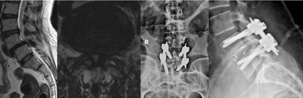
Patient with spondylolisthesis treated by MIS fusion
Minimally Invasive Cervical Foraminotomy
This is a MIS decompression procedure that enlarges the space in which a spinal nerve root exits the cervical spinal canal (intervertebral foramen). This narrowing can be caused by a herniated disc, bone spurs, thickened ligaments or joints, which may result in painful pinched nerves. The procedure is performed from the back (posterior) with the patient on his or her stomach.
A 1- to 2-cm incision is made on the symptomatic side of the neck. Using an operating microscope and x-ray guidance, the muscles are gradually dilated and a tubular retractor inserted to allow access to the cervical spine. Bone or disc material and/or thickened ligaments are then removed to decompress and relieve pressure on the spinal cord and/or nerves. The tubular retractor is removed, allowing the dilated muscles to come back together, and the incision is closed.
Vertebroplasty and Kyphoplasty
Vertebroplasty for the treatment of vertebral compression fractures (VCFs) was introduced in the United States in the early 1990s. The procedure is usually done on an outpatient basis, although some patients stay in the hospital overnight. The procedure may be performed with a local anesthetic and intravenous sedation or general anesthesia. Using x-ray guidance, a small needle containing specially formulated acrylic bone cement is injected into the collapsed vertebra. The cement hardens within minutes, strengthening and stabilizing the fractured vertebra. Most experts believe that pain relief is achieved through mechanical support and stability provided by the bone cement. Kyphoplasty, involves an added procedure performed before the cement is injected into the vertebra. First, two small incisions are made and a probe is placed into the vertebral space where the fracture is located. The bone is drilled and one balloon (called a bone tamp) is inserted on each side. The two balloons are then inflated with contrast medium (which are visualized using image guidance x-rays) until they expand to the desired height and removed. The spaces created by the balloons are then filled with the cement. Kyphoplasty has the added benefit of restoring height to the spine.

Patient with T11 compression fracture with severe instability back pain treated by vertebroplasty
Surgical Expertise
Testimonials
What Our Patients Say
EXCELLENT rating
Based on 107 reviews My father had rotator cuff problems and needed follow-up care.Dr.Ankith N V was exceptional. They provided thorough, compassionate care and clear communication. The clinic was well-equipped, and attentive Highly recommend.
 RAVINDRA KUMAR2024-06-09Very good Orthopaedic Clinic and Dr Ankith sir well experienced Dr and also clear all the doubts for patients and their families...
 Dilip Gowda BR2024-05-29I had an outstanding experience with Dr. Ankit at Ankit’s Orthopedic . Dr. Ankit’s exceptional friendliness and professionalism made me feel comfortable and well-cared for throughout my visit. I highly recommend Dr. Ankit for their exemplary patient care and welcoming demeanor.
 Nikhil L2024-05-29Doctor is so knowledgeable and humble. Also he listens to us keenly and understands the problem. Highly recommended.
 Akshay2024-05-29Good communicate with patients and cooperation well good, we are satisfied your treatment.
 renuka Mallikarjuna2024-05-22Best orthopaedic clinic in vijayanagar good service
 ಬಳ್ಳಾರಿ ನಾಗ2024-05-22Very good doctor and
 Kiran S2024-05-18Really...this clinic is too good i recommended Dr.Ankith sir ..and doctor treat very well, he had more knowledge and as well as good human being and his staff is wonderful, this is the perfect place for orthopedic and spine
 ashoka vc2024-05-17Good service well qualified doctor
 srinath v2024-05-17
Book Your Appointment Today!
Call us at 099644 83761 to schedule your visit. Your path to recovery begins at Dr. Ankith’s Orthopedic & Spine Care Clinic. We look forward to helping you achieve your health goals!