Cervical Laminectomy
- Home
- Cervical Laminectomy
Cervical Laminectomy
What is a cervical laminectomy?
Cervical spondylotic myelopathy (CSM) refers to inadequate function of the spinal cord due to compression by bony out growths (spondylosis), ossification of ligaments or by large disc herniations.
What causes spondylotic myelopathy?
Over time, the normal wear-and-tear effects of aging can lead to a narrowing of the spinal canal. This compresses — or squeezes — the spinal cord. CSM can cause a variety of symptoms, including pain, numbness, and weakness. Symptoms usually begin after the age of 50, but can occur earlier if there was an injury to the spine at a younger age. Many people will have steady progression of their disease. Once symptoms start, they tend to continue. Typically, the disease progresses slowly over several years
What are the symptoms?
The spinal cord serves as the conduit for transmission of signals between the brain and the rest of the body. When the spinal cord is slowly compressed, people may develop symptoms such as:
- Tingling and numbness in the hands and legs
- Weakness: difficulty in buttoning shirts, clumsiness, dropping things
- Difficulty walking (loss of balance), wide-based gait
- Coordination problems: changes in handwriting and feeding
- Neck pain and stiffness
What are the common investigations?
X-rays are useful to look at the alignment of the neck. Magnetic resonance imaging (MRI) is the ideal investigation to look for compression of the spinal cord. CT scans allow better detailed cross-section images showing bone spurs and the size of the spinal canal and are ordered in special situations.
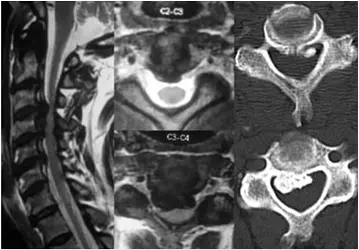
MRI and CT scan images showing extensive spinal canal compromise in a patient with myelopathy
What is the standard treatment?
Nonsurgical Treatment
In some people with early myelopathy without significant compression of the cord, medical management is initiated. Soft collars allow the muscles of the neck to rest and limit neck motion. This can help decrease pinching of nerve roots with movement. Soft collars should only be worn for short periods of time, because long-term wear can decrease the strength of neck muscles. Improving neck strength and flexibility with simple exercises may lessen discomfort. Nonsteroidal anti-inflammatory medications (NSAIDs) can reduce swelling and painful symptoms.
Surgical treatment
Surgical treatment includes anterior cervical decompression or posterior surgeries. Posterior decompression procedures include cervical laminectomy and cervical laminoplasty and are advised when there is multi-level compression of the spinal cord.
What is cervical laminectomy?
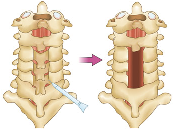
Picture showing back of the neck bones that has been treated by cervical laminectomy to increase the spinal canal dimension
When the spinal canal has become too small due to injury or disease, it may be made larger by use of laminectomy. By increasing the space for the spinal cord and nerve roots, laminectomy reduces the spinal cord compression and may help prevent progression of neurological deficits. An incision is made down the back of the neck to expose the cervical vertebrae. Then the laminas are cut all the way through on either side of its attachment to the vertebra. The lamina with the intervening ligaments is removed creating more room for the spinal cord and nerve roots.

MRI images before and after surgery indicate good spinal cord decompression after cervical laminectomy
What is cervical laminoplasty?
When the spinal canal has become too small due to injury or disease, it may be made larger by use of laminoplasty. By increasing the space for the spinal cord and nerve roots, laminoplasty reduces the spinal cord compression and may help prevent progression of neurological deficits. It preserves the stability of the neck. The lamina is a flat portion of bone that is the back portion of the vertebra. On one side of the vertebral column, the laminas are cut through just far enough to create a hinge-like movement, much like a door. Then the lamina on the other side are cut all the way through to, in effect, open the door. After gently opening the “door” of each vertebra to create more room for the spinal cord and nerve roots behind it, bone wedges are inserted to keep the “door” from totally closing. Then the “door” is closed securely onto bony wedges, resulting in an expanded “doorway” for the spinal cord and nerves.
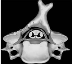
Axial view through the spinal canal showing 'widened' spinal canal after a laminoplasty
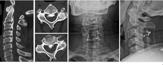
CT scan images show flowing ossification of ligamentum flavum treated with posterior cervical laminoplasty
Surgical Expertise
Testimonials
What Our Patients Say
EXCELLENT rating
Based on 107 reviews My father had rotator cuff problems and needed follow-up care.Dr.Ankith N V was exceptional. They provided thorough, compassionate care and clear communication. The clinic was well-equipped, and attentive Highly recommend.
 RAVINDRA KUMAR2024-06-09Very good Orthopaedic Clinic and Dr Ankith sir well experienced Dr and also clear all the doubts for patients and their families...
 Dilip Gowda BR2024-05-29I had an outstanding experience with Dr. Ankit at Ankit’s Orthopedic . Dr. Ankit’s exceptional friendliness and professionalism made me feel comfortable and well-cared for throughout my visit. I highly recommend Dr. Ankit for their exemplary patient care and welcoming demeanor.
 Nikhil L2024-05-29Doctor is so knowledgeable and humble. Also he listens to us keenly and understands the problem. Highly recommended.
 Akshay2024-05-29Good communicate with patients and cooperation well good, we are satisfied your treatment.
 renuka Mallikarjuna2024-05-22Best orthopaedic clinic in vijayanagar good service
 ಬಳ್ಳಾರಿ ನಾಗ2024-05-22Very good doctor and
 Kiran S2024-05-18Really...this clinic is too good i recommended Dr.Ankith sir ..and doctor treat very well, he had more knowledge and as well as good human being and his staff is wonderful, this is the perfect place for orthopedic and spine
 ashoka vc2024-05-17Good service well qualified doctor
 srinath v2024-05-17
Book Your Appointment Today!
Call us at 099644 83761 to schedule your visit. Your path to recovery begins at Dr. Ankith’s Orthopedic & Spine Care Clinic. We look forward to helping you achieve your health goals!